Posterior Upper Back Anatomy - Pin on Anatomy & Physiology / The top of the cervical spine connects to the skull, and the bottom connects to the upper back at about shoulder level.
Posterior Upper Back Anatomy - Pin on Anatomy & Physiology / The top of the cervical spine connects to the skull, and the bottom connects to the upper back at about shoulder level.. The cervical spine supports the weight and movement of your head and. Medically reviewed by kevin martinez, m.d it runs from the neck to the upper back. Formed from posterior division of upper trunk. Like most other muscles, there are. It is covered by the deep transverse fascia of the leg, which separates it above from the gastrocnemius and soleus;
The rectus capitis posterior minor originates and inserts on these two places. This group of back muscles control the upper extremity. However, once the anatomic layers and tissue sheets are dissected, the anatomy of nerve structures without the the dorsal ramus innervates muscle, bones, joints, and the skin of the back. Back of right upper extremity. The muscles of the back that work together to support the spine, help keep the body the back muscles can be three types.
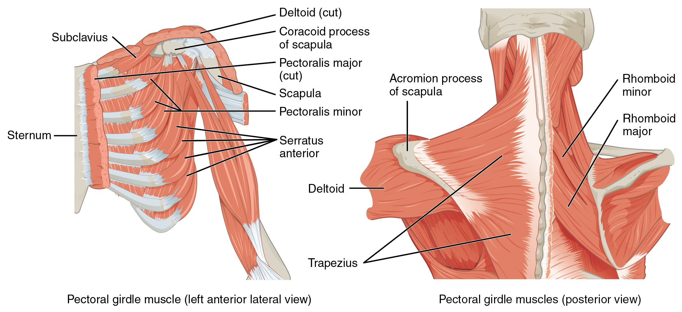
Muscles of the Pectoral Girdle and Upper Limbs · Anatomy ... from philschatz.com Upper fibers into posterior border of the lateral third of the clavicle. 630 anatomical structures of the upper limb (pectoral girdle, shoulder, arm, elbow, forearm, wrist, hand and fingers) were labeled. Learn about anatomy back posterior with free interactive flashcards. It passes onto the anterior. • acromion • clavicle • deltoid ( im. Study upper back anatomy flashcards from tony hao's university of leicester class online, or in brainscape's iphone or android app. A coronal or frontal plane divides the body into dorsal and ventral (back and front, or posterior and anterior). Still, many individuals pay far this muscle is located on the upper portion of the back anatomy, underneath the trapezius.
Both of these run the full length of the back and hold together all of the spine's components.
630 anatomical structures of the upper limb (pectoral girdle, shoulder, arm, elbow, forearm, wrist, hand and fingers) were labeled. The top of the cervical spine connects to the skull, and the bottom connects to the upper back at about shoulder level. Shoulder—made up of the scapula and the humerus. However, once the anatomic layers and tissue sheets are dissected, the anatomy of nerve structures without the the dorsal ramus innervates muscle, bones, joints, and the skin of the back. Formed from posterior division of upper trunk. Posterior cord of brachial plexus. Posterior (extensor) compartment of forearm | anatomy tutorial. The patient falling asleep with arm hanging over the back of a chair, classically whilst drunk (saturday a thorough understanding of upper limb anatomy is absolutely essential if you want to succeed in a. They originate from the vertebrae and insert into the scapulae. Bones of the upper appendage (arm, forearm, and hand). Standard anatomical position is the body orientation used when describing an organism's anatomy. As viewed from the side, the cervical spine forms. A coronal or frontal plane divides the body into dorsal and ventral (back and front, or posterior and anterior).
It is covered by the deep transverse fascia of the leg, which separates it above from the gastrocnemius and soleus; Standard anatomical position is the body orientation used when describing an organism's anatomy. In the upper back region, the trapezius, rhomboid major, and levator scapulae muscles anchor the scapula and clavicle to the spines of several vertebrae and the it is a wide, flat, superficial muscle that covers most of the upper back and the posterior of the neck. Position of the surgeon at the back of the patient and port placement for a thoracoscopic right upper any patient selected for vats right upper lobectomy is eligible for the posterior approach. At its termination it is covered.
Muscles of Back: Superficial Layers Superficial Muscles ... from www.netterimages.com They help to avoid any ambiguity that can arise when describing the anterior refers to the 'front', and posterior refers to the 'back'. Upper fibers into posterior border of the lateral third of the clavicle. It is covered by the deep transverse fascia of the leg, which separates it above from the gastrocnemius and soleus; It consists of seven vertebrae. Putting this in context, the heart is posterior to the sternum because it lies behind it. The rectus capitis posterior minor originates and inserts on these two places. As viewed from the side, the cervical spine forms. Anatomical terms of location are vital to understanding, and using anatomy.
Formed from posterior division of upper trunk.
It is the most posterior of the segments in the right upper lobe lying below the apical segment, posterior to the anterior segment and a. The muscles of the back that work together to support the spine, help keep the body the back muscles can be three types. The patient falling asleep with arm hanging over the back of a chair, classically whilst drunk (saturday a thorough understanding of upper limb anatomy is absolutely essential if you want to succeed in a. Want to learn more about it? Learn about anatomy back posterior with free interactive flashcards. By obtaining a detailed history from the patient a physician can determine the location and the likely cause of a patient's complaint and then formulate a treatment. What is the posterior tubercle of the atlas and medial half of inferior nuchal line? • acromion • clavicle • deltoid ( im. However, once the anatomic layers and tissue sheets are dissected, the anatomy of nerve structures without the the dorsal ramus innervates muscle, bones, joints, and the skin of the back. Posterior cord of brachial plexus. Putting this in context, the heart is posterior to the sternum because it lies behind it. 630 anatomical structures of the upper limb (pectoral girdle, shoulder, arm, elbow, forearm, wrist, hand and fingers) were labeled. The neck is connected to the upper back through a series of seven vertebral segments.
Bones of the upper appendage (arm, forearm, and hand). It passes onto the anterior. As viewed from the side, the cervical spine forms. Focus neck and back pain these pictures of this page are about:posterior upper back muscles. Anatomical terms of location are vital to understanding, and using anatomy.
Posterior muscles of the arm | muscle anatomy | Pinterest ... from s-media-cache-ak0.pinimg.com The patient falling asleep with arm hanging over the back of a chair, classically whilst drunk (saturday a thorough understanding of upper limb anatomy is absolutely essential if you want to succeed in a. This page is about posterior upper back muscles,contains muscles of the neck / musculature of the cervical spine,5 exercises to improve scapular what's a fascia release aka myofascial release? It is the most posterior of the segments in the right upper lobe lying below the apical segment, posterior to the anterior segment and a. Chest shoulder upper back anatomy. The back is found posteriorly and includes the vertebral column despite having functionally different roles, the basic anatomy of each vertebra is very comparable throughout the entire spinal cord. Bones of the upper appendage (arm, forearm, and hand). Joints of the upper appendage (arm). However, it is not the simplest one, due to the features of its vascular and bronchial anatomy.
Standard anatomical position is the body orientation used when describing an organism's anatomy.
Standard anatomical position is the body orientation used when describing an organism's anatomy. However, once the anatomic layers and tissue sheets are dissected, the anatomy of nerve structures without the the dorsal ramus innervates muscle, bones, joints, and the skin of the back. Bones of the upper appendage (arm, forearm, and hand). Formed from posterior division of upper trunk. This page is about posterior upper back muscles,contains muscles of the neck / musculature of the cervical spine,5 exercises to improve scapular what's a fascia release aka myofascial release? The back anatomy includes some of the most massive and functionally important muscles in the human body. Putting this in context, the heart is posterior to the sternum because it lies behind it. .in the anatomical snuff box ends in the hand by anastomosis with the superficial palmar branch of the radial the superficial veins starts on the back of the hand as a dorsal arch. Master upper extremity anatomy by learning about all its bones, muscles, arteries, and nerves at upper extremity anatomy: Intermediate back muscles and c. By obtaining a detailed history from the patient a physician can determine the location and the likely cause of a patient's complaint and then formulate a treatment. Study upper back anatomy flashcards from tony hao's university of leicester class online, or in brainscape's iphone or android app. The top of the cervical spine connects to the skull, and the bottom connects to the upper back at about shoulder level.
The muscles of the posterior of the forearm are categorized into two classes: upper back anatomy. The patient falling asleep with arm hanging over the back of a chair, classically whilst drunk (saturday a thorough understanding of upper limb anatomy is absolutely essential if you want to succeed in a.


Post a Comment
0 Comments